
How to protect yourself against breast cancer?
Breast cancer is the most common malignant tumor in women, accounting for over 20% of all cancer cases. Detecting the cancer at its earliest possible stage is crucial for successful treatment outcomes.
We talk about this dangerous disease, treatment options, and above all, prevention with Marcin Zając, a specialist in general and oncological surgery.
Doctor, how does breast cancer develop?
"The starting point for cancer development is a mutated process occurring in breast cells, which leads to their uncontrolled division and growth. Cancer cells, unlike normal cells, have lost the restraint of programmed cell death (apoptosis), have the ability to invade surrounding tissues, and form distant metastases. Breast cancer in most cases originates from cells lining the inside of the milk ducts. Breast cancer cells penetrate the lymphatic vessels and then reach the lymph nodes, causing them to enlarge. This is also a signal that the disease is becoming increasingly aggressive (breast cancer), and there is a greater likelihood that the cancer cells have also entered the bloodstream and could colonize other organs, causing metastasis.".
Can we list the main causes of breast cancer development?
– The etiology of breast cancer is not fully understood, despite numerous studies and analyses. The situation is further complicated by the fact that the same morphological tumor can be caused by several, or even a dozen, carcinogenic factors.
However, genetic factors are increasingly being implicated in the risk of developing breast cancer. Approximately 10% of breast cancers are hereditary. The likelihood of developing the disease increases with the number of first-degree relatives (mother, sister, daughter) with this cancer. Not all genes whose mutations can lead to breast cancer have yet been identified. The most reliable criterion for hereditary breast cancer is the detection of BRCA1 and BRCA2 . Women with BRCA have an 80% lifetime risk of developing breast cancer.
Other genes whose mutations may increase the risk of breast cancer are: ATP, BRIP1, TP3, CHEK and PTEN.
Other factors predisposing to the development of breast cancer include: age (the risk of developing breast cancer increases with age, and most cases occur in women over 50 years of age), early onset and late cessation of menstruation, late pregnancy (after the age of 35), and lifestyle.
It's known that the earlier cancer is detected, the greater the chance of recovery. How can changes in the early stages be recognized?
– The first symptom is a painless lump, usually located in the upper lateral part of the breast (approximately 35% of cases), and less frequently in the lower medial part. Other symptoms of breast cancer include nipple asymmetry, nipple retraction, ulceration of the nipple or breast skin, additional lumps in the area, unexplained pain, mild skin thickening, itching or burning of the nipple, and discharge from the nipple.
The developing cancer is often accompanied by enlargement of the lymph nodes in the armpit, and in later stages, enlargement of the cervico-supraclavicular nodes. Symptoms of so-called inflammatory breast cancer include rapidly increasing swelling of the skin, accompanied by redness, excessive warmth, and pain.
Women as young as 20 should develop the habit of breast self-examination. Standing in front of a mirror, raise your arms high in the air and examine the breasts for any changes in their shape, discoloration, wrinkles, or tightness. The same should be done with your hands on your hips. Then, check for any discharge by pressing on the nipple.
While in the shower, place your left hand behind your head and use your right hand to control your left breast. Using light pressure, use your three middle fingers to draw small circles along your breast, up and down, then back down. Repeat with your right breast.
Fluctuating hormone levels cause most women's breasts to become swollen and tender before and during their periods. The consistency of their tissue also changes slightly.
For this reason, breast examination (both self-examination, ultrasound or mammography) should be performed one week after your period.
The most important diagnostic methods for breast cancer include:
- mammography,
- ultrasound,
- biopsy,
- surgical excision,
- examination of nipple discharge cells.
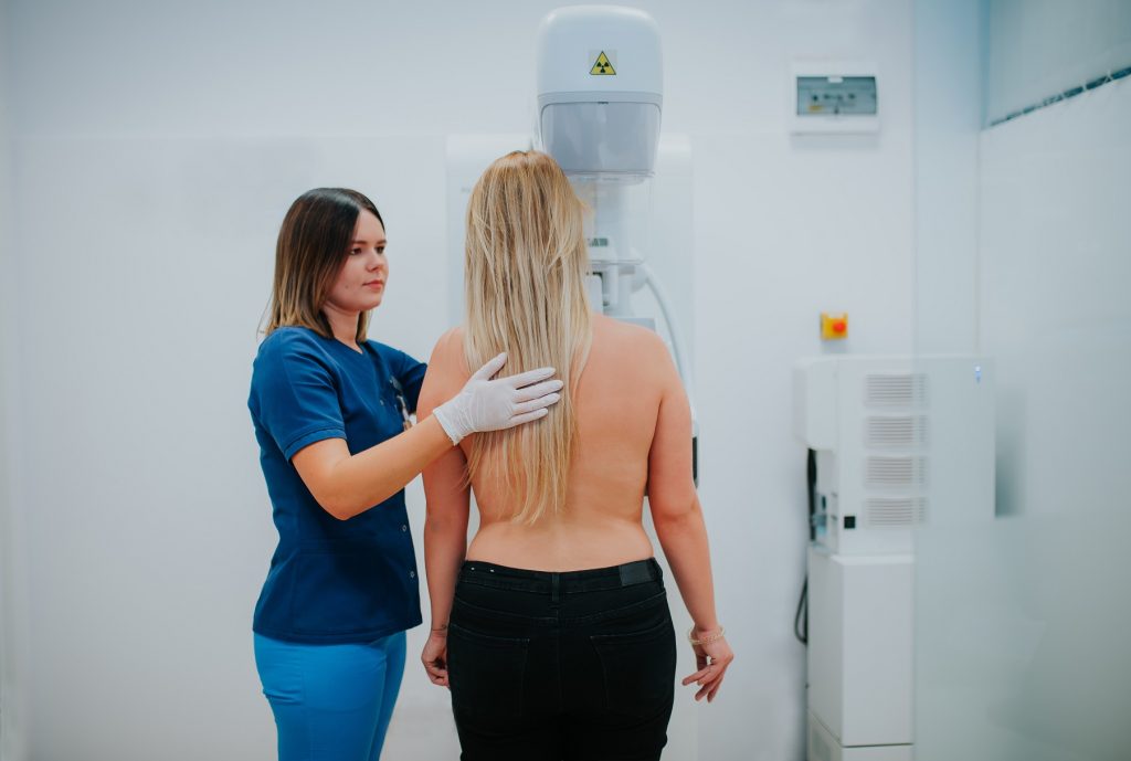
What exactly is a mammogram?
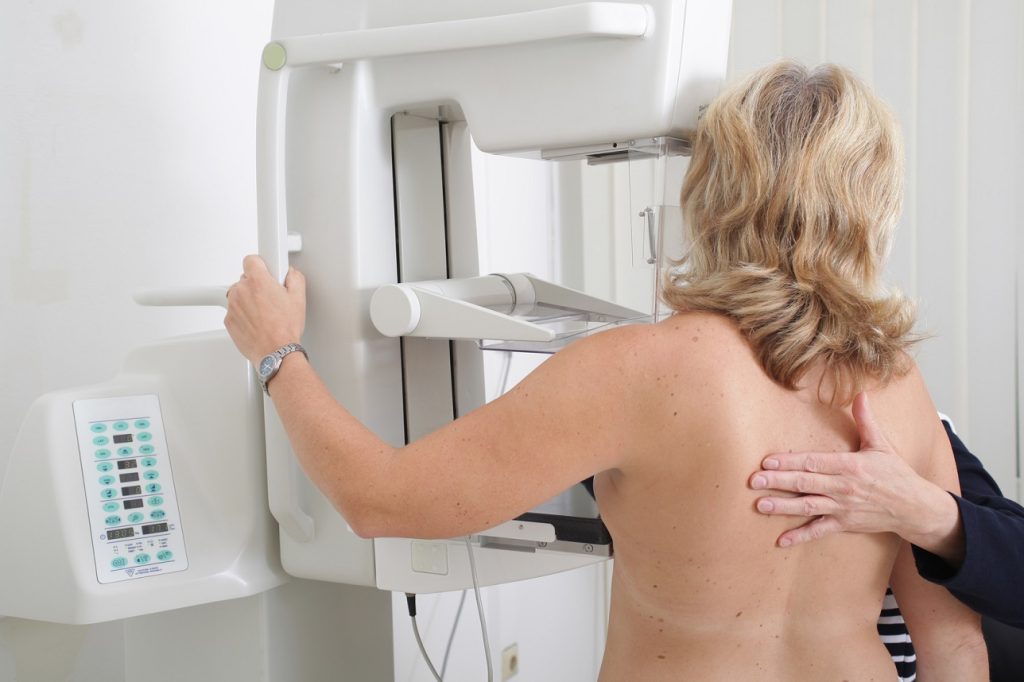
Currently, mammography is considered the best way to detect early breast cancer in women over 40. Mammography is an X-ray of the breast. This test can detect very small, unpalpable cancerous lesions. The sensitivity of mammography is estimated at 80-90%.
The examination is safe, the radiation during the examination is similar to the radiation during a dental X-ray.
Some women may experience some discomfort during the breast compression necessary for the examination.
Digital mammography is an extremely important step in the development of diagnostics. It significantly increases imaging precision, allows for analytical image transformation, and allows for the magnification of any fragment for more detailed analysis. The greater contrast range and the ability to adjust contrast are particularly useful in examining breasts with a large amount of glandular and fibrous tissue. Software image filtering not only improves image quality but can also aid diagnosis, for example, by automatically locating microcalcifications.
Mammography allows for determining the likelihood of cancer in a diagnosed lesion and selecting the appropriate further treatment. Mammographic findings should be classified by a radiologist according to the BIRADS (BreastImaging Reporting and Data System) system. Based on these results, the specialist/supervising physician will determine further recommendations and treatment.
The first mammogram is recommended around age 40 and should be performed every 1-2 years. For ages 50-69, screening is recommended every 2 years. After age 70, due to increasing life expectancy, regular screening is recommended until age 73.
But please note: if someone in your family has had breast cancer (mother, sister, daughter) or you have been diagnosed with mutations in the BRCA 1 or BRCA 2 genes, the frequency of the test should be discussed with your doctor.
There are special treatment protocols for this group of women (more frequent mammography, alternating with magnetic resonance imaging, and gynecological examination for ovarian cancer).
If the result is abnormal, the patient should consult an oncologist. The doctor will decide on further testing: guided mammography, ultrasound, or ultrasound-guided core or fine-needle biopsy.
What are the treatments for breast cancer?
Breast cancer treatment is a complex approach and should be performed in specialized oncology centers. Methods used include surgery, systemic therapy—chemotherapy, hormone therapy, targeted therapies, and radiotherapy.
Early detection of breast cancer allows for breast-conserving treatment, where only the lesion is removed, leaving the breast intact. This is the treatment of choice; if this is not feasible, the entire breast, including the lymph nodes and pectoral muscles, must be removed. In patients who require breast amputation, breast reconstruction can be performed.
Chemotherapy is based on pharmacological treatment, most often intravenously administered cytostatic drugs. It is used to destroy clinically undetectable micrometastases that may appear in the early stages of cancer, or to reduce the size of advanced tumors.
Radiotherapy – involves treatment with ionizing radiation.
It is often used as an integral part of sparing treatment.
Thank you for the interview.
For diagnostic tests, prior booking is required by calling 81 532 37 11 or online.
We collect laboratory tests (markers and basic tests) on an ongoing basis at collection points; we encourage you to purchase and pay online.