
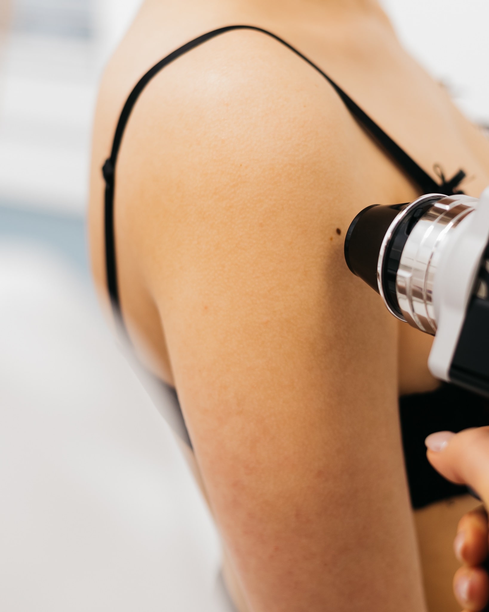
Videodermatoscopy - examination of moles, precise diagnosis
Videodermatoscopy is a non-invasive, painless, and highly accurate examination of moles and other skin lesions. It can be particularly useful when you notice pigmented lesions such as spots, birthmarks, or moles that have changed in a disturbing way, such as changing shape, color, or size. Such symptoms may indicate skin cancer, so skin care should be maintained year-round and routinely examined, and this is where videodermatoscopy comes in handy.
We talked about this innovative technology with dermatologist Joanna Nowak-Guzowska, who specializes in videodermatoscopy at CM Luxmed.
What is a videodermatoscope?
A video dermatoscope consists of a dermatoscope and an integrated digital camera with illumination. The image is captured and displayed on a monitor. A traditional handheld dermatoscope typically allows for a maximum magnification of 20x, while a video dermatoscope magnifies the lesion up to 400x. This allows the doctor to obtain an exceptionally detailed, high-definition image of the skin lesion.
Videodermatoscopy for examining moles and what else?
Videodermatoscopy is primarily helpful in diagnosing melanoma and precancerous lesions. It analyzes the appearance and type of various moles:
- pigmented, popularly known as moles,
- skin moles,
- lentigines and other discolorations,
- seborrheic keratosis,
- common warts,
- stellate hemangiomas,
- vascular malformations,
- ruby angiomas,
- hematomas on the skin and nails.
It is also helpful in diagnosing parasitic skin diseases (scabies, lice, demodicosis), as well as in diagnosing nail diseases and many other conditions such as psoriasis or lichen planus.
Why is early detection of skin melanoma important?
Early detection of skin melanoma is crucial because it significantly increases the chances of successful treatment and complete recovery, and also prevents the cancer from spreading to other parts of the body.
A key element of early melanoma detection is the preventive monitoring of skin lesions. Videodermatoscopy allows for the recording of moles and their comparison during follow-up visits, allowing us to identify and detect any changes in a given mole. Surgical excision can then be recommended at this early stage, before the mole develops into cancer.
Videodermatoscopy is a breakthrough in skin cancer prevention, especially melanoma. This advanced technology allows for high-quality images thanks to innovative software. It allows for the examination of moles at high magnification, and the resulting images can be precisely analyzed directly with the human eye, as well as using artificial intelligence, which is incorporated into the videodermatoscopy system's application.
This examination is particularly important in the case of moles located in hard-to-reach places, which are difficult for the patient to inspect on his own.
What does the examination look like and what are the contraindications?
The test is intended for everyone, regardless of gender, age, or skin type. After a thorough interview, the test begins with the doctor taking a localization photo (MACRO photo), which is a photograph of specific body parts. Next, the moles are marked in the app, and a camera (MICRO photo) is taken, but only of the selected moles. The images of the moles precisely depict their structure on the device's screen, which are then analyzed by the specialist.
As for contraindications, it is important that the skin is not tanned before the examination!
How long does a full body examination take?
The duration of a full-body examination depends on the number of skin lesions being examined. If we are analyzing single lesions (several moles), the visit may take 15-30 minutes. If the patient requires a detailed analysis of a larger number of moles located throughout the body, the examination may take up to one hour. After the examination, the patient receives recommendations for further treatment—removal or observation of individual lesions—as well as a detailed report with photos and descriptions.
Over the last 20 years, the incidence of melanoma has increased by as much as 300% and, unfortunately, this cancer is increasingly occurring in young patients aged 25-40.
Remember that early detection of melanoma allows for almost complete recovery. That's why prevention is so important. Videodermatoscopy allows patients to regularly monitor their moles, thereby diagnosing melanoma at the earliest possible stage.
If you notice disturbing skin changes, new moles or want to have your skin checked as a preventive measure, schedule a dermatological consultation with videodermatoscopy at CM Luxmed Lublin – Dermatology
Thank you for the interview
Frequently asked questions – FAQ
What is videodermatoscopy?
Videodermatoscopy is a non-invasive examination of skin and moles using an image-magnifying device. It allows for a precise assessment of the structure of a skin lesion and its digital documentation.
What is the difference between videodermatoscopy and dermatoscopy?
Dermatoscopy allows for the assessment of the lesion under magnification, and videodermatoscopy also allows for the recording of the lesion image, its comparison during subsequent visits, and more accurate monitoring of any changes over time.
When is it worth performing videodermatoscopy?
It is worth performing the test if you have numerous moles, new or changing skin lesions, family history, fair skin, frequent exposure to the sun, sunburn, or a disturbing appearance of a mole.
What signs should worry you?
Moles that are worrisome are those that grow rapidly, change color, shape or size, have irregular edges, are itchy, painful, bleed, are asymmetrical or clearly different from other skin lesions.
What does a videodermatoscopic examination look like?
The doctor examines the skin and selected lesions using a video dermatoscope, which provides a highly magnified image. If necessary, photographic documentation is taken for comparison during subsequent checkups.
Does videodermatoscopy hurt?
No. Videodermatoscopy is a painless and non-invasive examination. It does not require skin punctures, anesthesia, or a recovery period.
How to prepare for videodermatoscopy?
Before the examination, it's a good idea not to apply makeup or cosmetics to the areas being assessed. It's also a good idea to remind yourself of any moles that have changed in appearance or have appeared recently.
How often should you check your moles?
The frequency of checkups depends on the number of moles, skin type, family history, previous sunburns, and the assessment of a dermatologist. For many people, checkups are recommended once a year, or more frequently as risk changes.